As an advanced manufacturing technology integrating light/machine/electricity, computer, numerical control and new materials, 3D printing technology has been widely used in aerospace, military and weapons, automotive and racing, electronics, biomedicine, dentistry, jewelry. Games, consumer goods and daily necessities, food, construction, education and many other fields are now a trend of rapid development. In theory, all materials can be used for printing. For the high-end sector, the limitations of printed materials have seriously hampered the development of printing technology. The bottleneck of printed materials has become one of the key issues in the study of 3D printing.
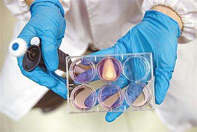
Yang Yizhen, chairman of Shenzhen Guanghua Weiye, believes that the current problems of 3D printing materials are mainly reflected in the following points: the maturity of applicable materials cannot keep up with the development of the printing market; the fluency of material printing is not enough; the strength of special materials cannot meet the requirements; Safety and environmental friendliness; material standardization and serial management issues. Solving a series of problems with printed materials is particularly important, directly related to whether 3D printing technology can lead us into a new era of rapid manufacturing. Among the materials studied in biomedical applications are the most eye-catching, because the materials in this area are the most difficult to do and the most expensive. 3D printing of biomedical materials is particularly difficult, considering the strength, safety, biocompatibility, degradability of tissue engineering materials, etc. The biomedical materials currently available for 3D printing mainly include metals, ceramics, polymers, Bio-ink, etc., is characterized by a wide range of distribution, but few types. At the beginning of 2017, the Antarctic bear learned that after implanting 3D printed blood vessels into monkeys, Blu-ray Yinguo plans to start clinical implantation in human body in 2017.
Application of 1.3D printing technology in biomedical engineering
At present, 3D printing technology is widely used in the field of biomedicine, including not only the physical manufacture of bones, teeth, artificial liver, artificial blood vessels, pharmaceutical manufacturing, etc., but also the international use of this technology for the manufacture of organ models and surgical analysis planning. , the manufacture of personalized tissue engineering scaffold materials and prosthetic implants, as well as applications in cell or tissue printing. According to reports, in December 2013, the Institute of Regenerative Medicine of the University of Cambridge pioneered the use of 3D printing technology to prepare artificial retinas with three-dimensional structure using ganglion cells and glial cells of rat retina. The artificial retinal cells have a high survival rate after printing and still have the ability to divide and grow. This breakthrough progress has brought hope to human cure for blindness. Some cell-free repair materials have been prepared using 3D printing technology and biomimetic materials, and have been applied clinically. In the future, 3D printing technology can be used to print biologically active human organs to realize the clinical application of artificial organs. In addition, 3D printing technology can be used for personalized treatment, reducing treatment costs, and developing more biocompatible and biodegradable materials in the future. Combined with 3D printing technology, it can reduce the damage caused to the human body due to lack of materials. In this way, 3D printing technology will lead the revolutionary trend in the medical field.
2.3D printing biomedical materials
2.1 medical metal materials are currently used for research
3D printed biomedical materials are mostly plastic, and metal materials have better mechanical strength, electrical conductivity and ductility than plastics, making them have natural advantages in the field of hard tissue repair research. The melting temperature of metal is relatively high, and printing is difficult. Therefore, metal 3D printing is generally processed by photo-curing 3D printing (SLA) and selective laser sintering (SLS), which is produced by irradiation of metal powder under ultraviolet light or high-energy laser. The high temperature achieves the fusion of the metal powder and superimposes layer by layer to obtain the desired components. Currently, SAHOD materials for biomedical printing mainly include titanium alloys, cobalt chromium alloys, stainless steels, and aluminum alloys. The team led by Prof. Guo Zheng, Department of Bone Oncology, Xijing Orthopaedic Hospital, Xi'an Fourth Military Medical University, used metal 3D printing technology to print a titanium alloy implant prosthesis that is completely consistent with the patient's clavicle and scapula, and successfully implanted titanium alloy prosthesis through surgery. In patients with bone tumors, it has become the first application of reconstruction of the scapular zone in the world, marking the further maturity of 3D printing individualized metal skeleton repair technology.
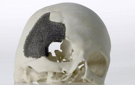
Compared with the traditional individualized implant prosthesis preparation technology, the 3D printed individualized titanium implant prosthesis of clavicle, scapula and other amorphous bone has higher matching, and the function and shape are more recognized by patients and doctors; Porous design stone bone and soft tissue attachment aging rate is high; elastic modulus is reduced, reducing stress shielding complications; product quality is stable, accuracy can reach 1mm; short preparation cycle and other advantages. At present, the shortcoming of this technology is that the printing material is expensive, and the patient needs to bear a large economic burden, and it is difficult to achieve civilianization. The Institute of Physics and Chemistry of the Chinese Academy of Sciences uses low-melting-point metal 3D printing technology, such as liquid metal Ga67In20.5Sn12.5 alloy (melting point is about 11 ° C), combined with minimally invasive surgery to directly inject the medical electronic device at the target tissue in the living body. Innovative research. They first inject a biocompatible encapsulating material (such as gelatin) into a biological tissue to form a specific structure, and then use a tool (such as an injection needle) to pierce and pull out in the cured package area to form an electrode area, and finally to conduct electricity. Metal inks, insulating inks, and even micro/nanoscale devices are sequentially injected to form target electronic devices. By controlling the 3D printing step of the microinjector's needle insertion direction, injection site, injection amount, needle displacement and speed, the terminal device can be constructed in a predetermined shape and function at the target tissue. They used this technology to prepare 3D liquid metal REID antennas in living tissues. The flexible devices fabricated by this in vivo 3D printing technology have high compliance, conformity, and minimally invasive and low-cost features. It shows a good application prospect and is of great significance in the field of implantable biomedical electronics.
With the advent and development of nano 3D printing technology, nano-powder printing materials have become a hot topic for researchers, and metal powder has occupied the main position in the 3D printing powder market. Advanced nanostructured powders have high requirements for ultra-fine crystal structures. Nanostructured powders can significantly improve the physico-chemical properties of printed products. These performance enhancements will further broaden their application in biomedical applications. However, the industrialization and commercialization of these nanopowders is still very difficult due to processing difficulties, low production efficiency, and high cost.
2.2 medical inorganic non-metallic materials
Inorganic non-metallic biomaterials mainly include bioceramics, bioglass, oxide and calcium phosphate ceramics and medical carbon materials. Among them, bioceramics have excellent properties such as high hardness, high strength, low density, high temperature resistance and corrosion resistance, and have wide applications in medical bone substitutes, implants, dental and orthopedic prostheses. However, the toughness of bioceramics is not high, and the hard and brittle characteristics make it difficult to form and shape. Especially the ceramic parts with complex shape or internal structure need to be formed by the mold, and the mold processing is expensive and the development cycle is long, which is difficult to meet the demand of the product. In recent years, researchers have made great progress in the preparation of bioceramics using 3D printing technology for the problems of complex bioceramic manufacturing processes and difficult molding processes.
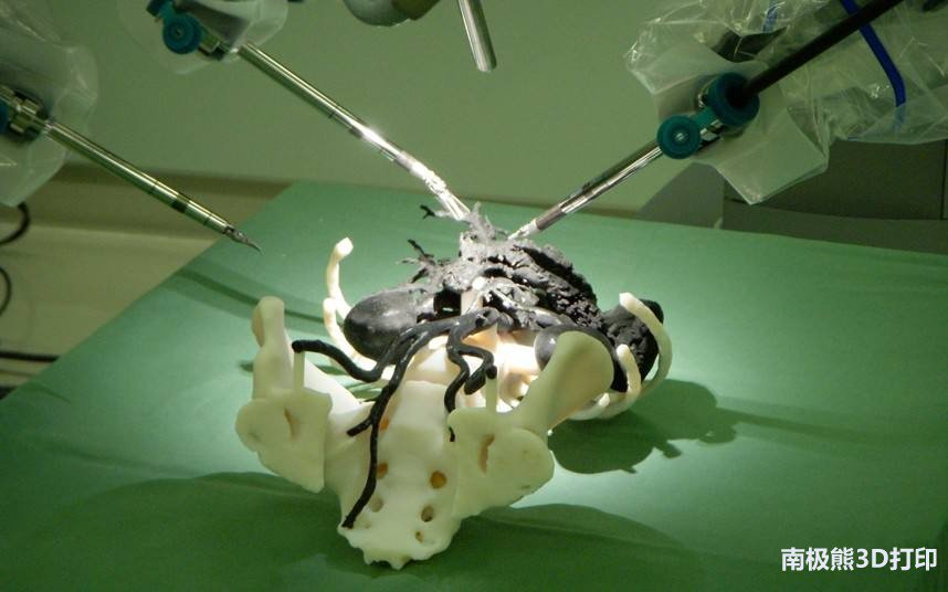
Saijo and other biomaterials such as tricalcium phosphate powder are used to prepare personalized prosthesis. After treatment, it can be directly implanted into the human body without engraving during surgery. 3D printing is introduced into the field of cosmetic surgery and has achieved good results. The use of 3D printing technology to manufacture cosmetic plastic materials can not only achieve various personalization requirements of customers, but also achieve one-time precision molding, which eliminates the cumbersome pre-operative engraving process of traditional techniques, which greatly saves operation time and thus is widely used. attention. At present, there are mainly bioceramics such as calcium phosphate, calcium dibasic calcium phosphate, biphasic calcium phosphate, calcium silicate/β-tricalcium phosphate. The 3D printed ceramic scaffold has the biological activity function of promoting osteogenic differentiation and angiogenesis of cells. The hydroxyapatite scaffold can promote the differentiation of sphincter stem cells into osteoblasts, and the content of β-tricalcium phosphate in the biphasic calcium phosphate scaffold Increased, the ability of the scaffold to promote osteogenic differentiation of cells, the release of silicon in the calcium silicate/β-tricalcium phosphate scaffold can promote the synthesis of osteoblasts into osteogenic factors and promote osteogenic differentiation of cells. Calcium diorton phosphate promotes proliferation and regeneration of blood vessels. Bioceramics have similar compressive strength and good osteoinductive ability to cancellous bone, but bioceramics need to be printed in a high-temperature environment, and bioactive molecules or anti-infective drugs that promote bone formation can not be applied to the stent simultaneously. At the same time, it has high brittleness, poor toughness and weak shear stress. Current 3D printing studies on bioceramics are limited to hard tissue printing. Bioglass is an aggregate of silicates in which the internal molecules are randomly arranged. It mainly contains several metal ions such as sodium, calcium and phosphorus. Under certain ratios and chemical reaction conditions, a complex containing calcium hydroxyphosphate is formed. It has high biomimeticity and is the main inorganic component of biological bone tissue.
Bio-glass materials have the degradability and biological activity and can induce the regeneration of bone tissue. Therefore, they are widely used as tissue engineering scaffold materials in the research field of bone tissue engineering, and have a very broad application prospect in the field of inorganic non-metal materials. The researchers used bioglass materials to prepare the monkey thigh bone, implanted in the body, and after a certain period of time, the study found that the regenerated monkey bone cells have grown into the network structure of the bioglass, and the combination is very tight; Mechanical experiments have found that this artificial bone has better mechanical properties than the original bone. In 2011, researchers at Washington State University in the United States used 3D printing technology to print calcium phosphate into a skeleton-like structure that can be used as a support for new bone growth before decomposition. It has been successfully tested in animals. Satisfactory results. The biocompatibility of bioglass combined with 3D printing precision molding, rapid manufacturing, personalization and many other advantages will definitely make new breakthroughs in tissue engineering scaffolding materials and personalized medical fields.
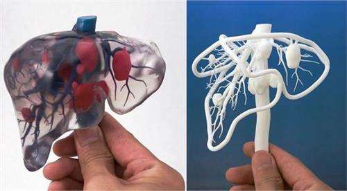
Since the above medical ceramic materials need to be processed under high temperature conditions, the 3D printing process of medical ceramic materials is usually divided into two stages:
1. The ceramic powder is uniformly mixed with the binder with lower melting point, and the designed model is sintered under laser irradiation. However, the model at this time only bonds the ceramic powder under the action of the binder, and the mechanical properties are poor. Unable to meet application requirements;
2. After laser sintering, the ceramic product needs to be placed in a muffle furnace for secondary sintering. The thickness of ceramic powder and the amount of binder will affect the performance of ceramic products. The finer the ceramic powder, the better the grain growth during secondary sintering. The better the quality of the ceramic layer; the larger the amount of binder, the laser sintering The easier the process is, but it will cause the parts to shrink when the secondary sintering is performed, so that the product does not meet the dimensional accuracy requirements. The temperature control of the secondary sintering process also has an effect on the performance of the 3D printed ceramic article.
2.3 medical polymer materials
In recent years, biomedical polymer materials have emerged as the fastest growing biomedical materials. The development of biomedical polymer materials has been designed from the very beginning by using only ready-made polymers to synthesize and synthesize polymers with special functions at the molecular level. At present, the research has entered a new stage, and it is a hot spot to find a kind of bioactive materials with active induction and stimulation of tissue regeneration and repair. 3D printing polymer consumables need special treatment, and also need to add adhesive or photocuring agent, and have high requirements on the curing speed and curing shrinkage rate of the material. Different printing technologies have different material requirements, but both require rapid and accurate material forming process. Whether the material can be quickly and accurately formed is directly related to the success or failure of printing. Since biomedical materials directly interact with biological systems, biomedical materials must have good biocompatibility in addition to various physical and chemical properties. The development of biomedical materials has stricter audit procedures than the development of other functional materials, so Research on 3D printed polymer materials for biomedical applications is just beginning.
Cho, etc., from Pohang University of Science and Technology in South Korea, uses PPF as a raw material, and the porous scaffold prepared by using photo-curing stereoscopic printing technology (SLA) has mechanical properties similar to those of human cancellous bone, and the scaffold can promote adhesion and differentiation of fibroblasts. In addition, the results of the transplantation of the PPF scaffold into the subcutaneous or skull defect of the rabbit showed that the PPF scaffold caused mild soft tissue and hard tissue response in the animal, such as inflammatory cells, angiogenesis and connective tissue after 2 weeks of transplantation. Formation, after 8 weeks, the inflammatory cell density decreased and more regular connective tissue was formed. Compared with traditional tissue engineering scaffolds, 3D printed tissue engineering scaffolds can be freely designed in shape, size, pore structure and porosity. Researchers can select the scaffold structure to be printed according to the repair requirements of different tissues. Paulius Danilevicius and others successfully prepared three-dimensional porous polylactic acid (PLA) tissue engineering scaffolds by laser three-dimensional printing technology, and carried out a series of studies on the effects of scaffold porosity on cell adhesion, growth and reproduction.
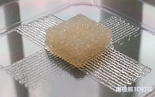
The results show that in the process of preparing the stent model, the three-dimensional printing technology can freely manufacture PLA tissue engineering scaffolds with arbitrary voids and porosity, and the researchers can easily obtain the required models. After a series of cell biological characteristics were characterized by various models, it was found that the cavity and porosity of the scaffold had a great influence on the adhesion growth of the cells. After analyzing and comparing the results, the most suitable model for tissue engineering scaffolds was obtained. . It has also been proved that PLA stents prepared by 3D printing are expected to be widely used in bone tissue engineering. Medical polymer printing materials have excellent processing properties and can be applied to a variety of printing modes, the most widely used are fused deposition printing and UV curing printing. The fused deposition printing uses thermoplastic polymer materials. Currently, the most popular researchers are degradable aliphatic polyester materials such as PLA and PCL. The raw materials only need to be drawn into a filament to print, and the preparation process of the printed materials is simple, and generally no need to add a printing aid. UV curing printing uses a liquid photosensitive resin containing a polymer monomer, a prepolymer, a light (sensitizing) curing agent, a diluent, etc., and the composition of the liquid resin and the degree of photocuring affect the printed product. Performance, especially biocompatibility and biological activity of medical products.
2.4 composite biomaterials
Composite material refers to a material composed of two or more different physical structures or different chemical properties, which are combined in microscopic or macroscopic form; or a multiphase material in which a matrix of a continuous phase and a reinforcing material of a dispersed phase are combined. It is called a composite biomaterial in the field of health care such as artificial organs, repair, physical therapy rehabilitation, diagnosis, examination, and treatment of diseases, and has good biocompatibility. FalguniPati et al. successfully printed PCL/PLA/β-TCP composite biomaterial scaffolds using multi-nozzle 3DP technology, and planted hTMSCs cells in scaffolds for 2 weeks to attach the extracellular matrix secreted during hTMSCs cell growth to the scaffold. Then, decellularization experiments were performed to remove the hTMSCs cells on the scaffold, and the extracellular matrix was retained, thereby obtaining a PCL/PLA/β-TCP/ECM multi-component bioactive composite biomaterial scaffold. The material in the stent can be well taken from the human body, the shortness of the supplement, and the components complement each other, which can not only meet the mechanical requirements of the bone tissue engineering material, but also promote the biomineralization process. ECM also contains a variety of factors that regulate the growth and differentiation of bone cells, and is expected to become a new direction in the study of bone tissue engineering scaffold materials.
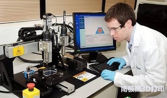
At the same time, FalguniPati et al also carried out research on 3D adipose tissue engineering. The first group used PCL as the framework to print a three-dimensional acellular fat scaffold with certain shape and pores in the PCL frame with decellularized adipose tissue as ink. Into the mice; the second group directly used the decellularized adipose tissue to load the target cells to make a gel, and the gel was printed in a previously prepared PCL frame by 3D printing technology, and the mice were implanted in vitro for a period of time. in vivo. Studies have shown that the tissue engineering scaffolds prepared by these two methods have good biocompatibility and can grow the required adipose tissue in mice. In general, the test data of the second group are all Better than the first group. It can be seen that 3D printing technology can print a variety of materials in combination, and each component can complement each other and complement each other, which has unique advantages in the field of tissue engineering. The properties of the composite biomaterial are tunable compared to single component or structural biomaterials. Since a single biomaterial can be made into a product by 3D printing, two or more biomaterials can be organically compounded together, and the components of the composite material maintain the relative independence of performance and complement each other. Configuration, greatly improving the shortcomings in the application of single materials; but for the two materials with different physical and chemical properties, how to use the printing method to integrate them well, to maximize the advantages of their combination is also the current research hotspot one.
2.5 cell-involved bio-3D printed materials
As a preliminary study, scientists have attempted to co-culture with cells using a number of 3D print holders, demonstrating that cells can survive on a variety of 3D print holders and perform better than normal two-dimensional cultures. The 3D printed PCL scaffold has been proven to be co-cultured with a variety of cells, which provides a good basis for mixing cells and materials into "bio-ink" and co-printing biological tissue. But this is only a two-dimensional effect of cells and materials, and does not directly place cells in the printing system, can only be called biological 3D printing that is not directly involved in cells. Biological 3D printing, in which cells directly participate, is a multidisciplinary and cross-disciplinary superdisciplinary that requires the use of principles and techniques in many disciplines such as biology, medicine, materials science, computer science, molecular biology, and biochemistry. The choice is one of the difficult points that need to be broken. Hydrogel is a multi-component system composed of a three-dimensional crosslinked network structure and a medium of a polymer, and has attracted wide attention as a new type of biomedical material. Medical hydrogels have good biocompatibility, their properties are similar to those of extracellular matrix, and their ability to adhere to proteins and cells is weak, which does not affect the normal metabolic process of cells. The presence of a hydrogel allows for cell protection, cell-to-cell adhesion, and organ configuration.
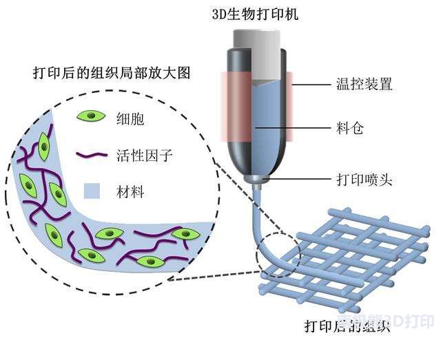
Therefore, hydrogels are the first choice for wrapping cells. Medical hydrogels, biocrosslinkers (methods), and living cells together constitute the "bio-ink" required for bio-3D printing. Researchers at Cornell University in the United States used 3D bioprinting technology to successfully print out the human auricle using "bio-ink" composed of type I collagen hydrogel and bovine ear cells. Whether it is function or appearance, this auricle is very similar to the normal human ear. During the subsequent culture, the collagen hydrogel interacts well with the cells and slowly degrades during the culture and is replaced by the extracellular matrix synthesized by the cells themselves. Next, they will use the patient's own ear cells to print the artificial auricle and transplant it. This news has created an infinite imagination for the future of the medical and plastic surgery industry.
The artificial auricles currently used in the medical profession are mainly divided into two categories:
One is made of artificial material like cartilage, and its disadvantage is that the texture is quite different from the human ear;
The second is to "carve" the new auricle by taking out part of the rib cartilage of the patient. This method not only causes a lot of physical damage to the patient, but also its aesthetic and practical degree is seriously controlled by the doctor's "engraving technique".
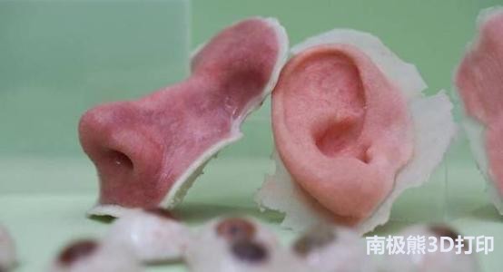
The artificial auricle made by 3D bio-printing technology does not have the above-mentioned flaws. Organ 3D printing is one of the dreams that scientists have been pursuing. At present, organ printing has been listed as a stock market speculation, which has attracted a lot of attention, but 3D printing is still in its infancy, and there are still many problems to be solved, especially complex organs. The 3D printing presents a greater challenge. The compatibility of materials with regulatory cells, the internal vascular construction of organs, and the growth factors of nervous system construction are the most difficult problems for organ printing. Biocompatibility cells, scaffold materials, growth factors, signal molecules, etc. are printed on the lower layers of computer instructions through 3D printing equipment to form living organs with physiological functions, which can be repaired or replaced. It is extremely widely used in biomedical fields. Use and prospects. In recent years, 3D printing technology has developed rapidly, and has achieved some achievements in bone, blood vessel, liver, breast construction, etc., but there is still a long way to go before the function of complex organs is realized.
to sum up
The development of 3D printing technology has become an emerging technology, and its application in medicine has become increasingly obvious. First, 3D printing technology will overcome the difficulties of tissue damage and organ failure. Everyone's own tissue and organs can be played at any time, which is equivalent to establishing their own tissue organ storage system for everyone. Secondly, the level of skin repair and beauty application will be further improved. As print accuracy and material adaptability improve, all parts of the body will be finely trimmed and blended to help create a more aesthetically pleasing human body. Finally, when 3D printing devices are gradually popularized, in some emergency situations, 3D printers can be used to make medical supplies, such as catheters and surgical tools, to make them more personalized, while reducing access and time, and temporarily solving medical supplies. The problem.
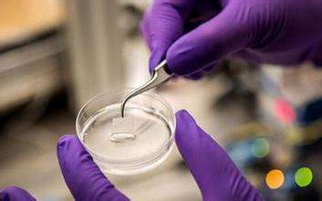
Therefore, the future development trend of 3D printing technology will increase the speed of 3D printing, develop more diverse 3D printing materials, make 3D printers smaller in size, lower in cost, and expand the application of more industries. At present, 3D printing has been mushrooming in biomedical research. 3D printing technology has made many achievements in the preparation of biomedical materials, especially tissue engineering scaffold materials. However, 3D printing biomedical materials are still a very fresh field, and various studies are still in the initial stage. There is still a long way to go to realize the clinical application of 3D printing biomedical materials. There are still great challenges. . The research and development of materials restricts the development of 3D printing technology, and the research and development of biomedical materials suitable for 3D printing will become a hot research topic in the future. The main reason for the difficulty in the development of 3D printing biomedical materials is that they have extremely high requirements on the various properties of materials in clinical practice. The choice of materials is restricted by many factors, considering the safety of materials before and after printing. Biocompatibility, degradation performance, bio-responsiveness, etc., must also consider whether the material can meet the requirements of industrialization. Therefore, the development of 3D printing biomedical materials faces enormous challenges. As the 3D printing technology develops rapidly in terms of programs and machinery, there are also many opportunities. Future research on 3D printed biomedical materials should focus on developing more printable biomaterials.
In theory, all materials can be printed, but in fact there are only a handful of printed materials for biomedical applications. Some materials with excellent properties are excluded from the ranks of 3D printed biomaterials due to the large shrinkage rate before and after printing, the additives contained in the materials are harmful to the organism, and the strength after printing is degraded, which cannot meet the requirements for the use of biomaterials. . Therefore, various physical and chemical modification methods should be used to overcome the printing problems of these discarded materials, and to develop more excellent 3D printed biomaterials. This can increase the choice of clinical application and reduce printing costs to a certain extent. Research and development of tissue engineering scaffold materials. 3D printing technology can design the spatial structure of the printing product arbitrarily. Combining this advantage of 3D printing with the organizational engineering concept, you can design the optimal tissue engineering bracket for a specific organization. In terms of material selection, the material that is closer to the extracellular matrix is ​​favored. Therefore, more biomimetic, biodegradable, bioactive 3D printed tissue engineering scaffold materials need to be developed.
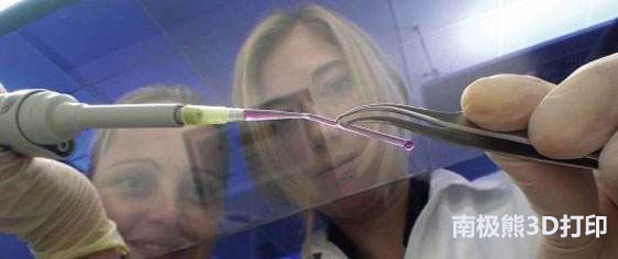
The combination of 3D technology and tissue engineering will open up new areas of research for the reconstruction of biological tissues and organs. Research and development of 3D printed biomaterials in "Bio Ink". Achieving in-situ 3D printing of tissues and organs is the result of scientists' dreams. The current state of the art only achieves the printing of materials and organs with invisible shape in vitro, and printing materials are one of the difficulties. Developed a bio-printing material with appropriate mechanical properties, good biocompatibility and biological activity, and composed it with living cells, bio-crosslinkers (methods) and signal molecules to form "bio-ink", striving to present the current 3D printing organs. Many problems have been solved, laying the foundation for realizing the benefits of 3D printing. In addition, research on the compatibility of printed materials with cells, tissues and blood is also one of the focuses. With the development of materials science, the requirements for bio-printing materials are becoming more and more stringent. The printed materials are not only safe and non-toxic, but also play the role of a scaffold. They also require certain biological functions to ensure the free exchange of material energy. , cell activity and three-dimensional construction of tissues. Therefore, research on the biocompatibility of printed materials is indispensable.
Author: Luo Wenfeng, Yang Xuexiang, Ao Jian Ning (Department of Biomedical Engineering, Jinan University, Guangdong Provincial Department of Education Key Laboratory of Biomaterials)
(Editor)
Facial Napkins,Kleenex Car Tissues,Great Value Facial Tissue,Bamboo Facial Tissue
Baoda Paper Enterprise Co., Ltd. , https://www.baodatissue.com